Share Post
Detecting Arthritis: Can X-rays Reveal the Condition?
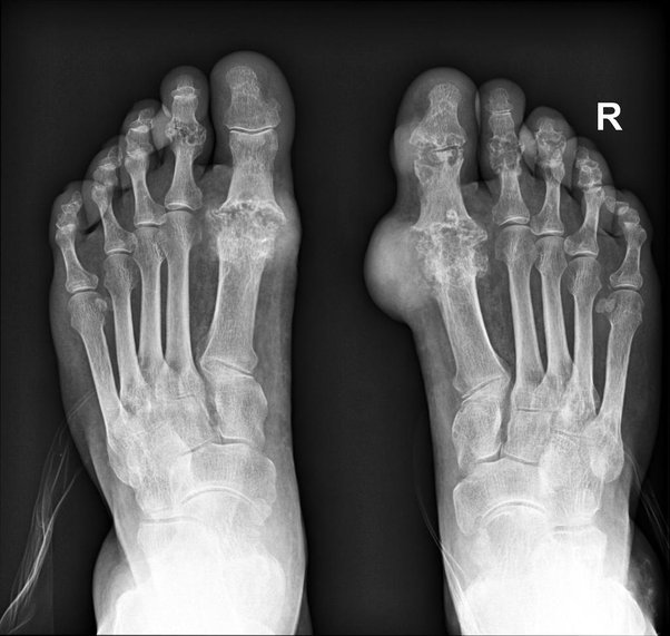
X-rays are frequently used to identify and track the progression of arthritis because they can reveal important details regarding changes to the bones and joints. An X-ray scan can show the degeneration and inflammation of the joints that are a feature of arthritis. Is this bad news, though? Normally no.
A narrowing of the joint space, bony spurs or osteophytes, changes in bone density, and the presence of cysts or erosions in the affected joint can all be signs of arthritis on an X-ray.
Every person experiences joint wear and strain. Osteoarthritis is the kind, which develops as we age. In fact, 90% of adults over the age of 40 who had their X-rays looked at would have osteoarthritis symptoms. Only a small portion of this 90% would, however, admit to experiencing any discomfort or stiffness at these joints. X-rays are usually used to diagnose arthritis, with osteoarthritis (OA) being the most prevalent type. OA is associated with genetic factors, wear-and-tear processes, and injuries, and is a natural aspect of aging.
An X-ray finding is only unusual if it develops earlier than it should or if it is an inflammatory form, such as rheumatoid arthritis or spondylitis.
There are other types of medical imaging that can help with the diagnosis of arthritis. In actuality, more effective methods exist, such as MRI and CT.
Early in the course of the disease, x-rays may appear normal with subtle components that make a diagnosis challenging. In certain situations, more advanced imaging, particularly MRI, may be required. In addition to soft tissue problems linked to arthritis, an MRI can frequently reveal earlier alterations in the bones.
In addition to the structural changes in the bones and joints, X-rays can also reveal certain soft tissue anomalies associated with arthritis. These may include:
- inflammation or thickening of the joint capsule
- Bursitis is an inflammation of the fluid-filled sacs (bursae) that cushion the joints. X-rays can assist detect symptoms of bursitis. The presence of fluid buildup or thickening around the afflicted joint is a sign of bursitis.
- Edema of the bone marrow (Swelling)
- Tendon involvement: X-rays may show abnormalities, such as thickening, calcification, or erosion, in the tendons close to the joint, which can happen in some kinds of arthritis.
X-rays can be performed quickly and simply. Additionally, they are useful in treating problems including fractures and serious misalignments. Your best bet is a thorough subjective history and an expert physical examination.
Although X-rays might reveal structural changes linked to arthritis, it’s important to keep in mind that these changes might not always be correlated with the severity of symptoms or the degree of joint destruction. To offer a more thorough evaluation of the joint and surrounding tissues, further imaging modalities like MRI or ultrasound may occasionally be used. The X-ray data will be interpreted by a healthcare professional, such as an orthopedic or rheumatologist, who will also take into account other clinical aspects to establish an accurate diagnosis and choose the best course of action.
A visit to Feldman & Leavitt Foot And Ankle Specialists will CLEARLY define all available patient options.
As a general rule, procedures are performed on an outpatient basis in an Alberta Health Services (AHS) approved Surgical Center or in a Hospital. Surgical procedural costs are covered by AHS or the patient may opt for private surgery to avoid a waiting time.
Schedule an Appointment now at Feldman & Leavitt Foot and Ankle Specialists, your expert podiatrists in Calgary, Alberta and the surrounding areas.